Research
Here are some examples of peer-reviewed, published manuscripts that highlight important new techniques and protocols in development at
T Lab that will improve detection of vector-borne pathogens and facilitate understanding of how related diseases develop.
Novel testing methods reveal vector-borne pathogen previously not detected.
The diagnostic tests available to identify vector-borne pathogens have major limitations. Clinicians must consider an assortment of often diverse symptoms to decide what pathogen or pathogens to suspect and test for. Even then, there are limitations to the currently available indirect detection methods, such as serology, or direct detection methods such as molecular tests with or without culture enrichment. Bartonella spp., which are considered stealth pathogens, are particularly difficult to detect and diagnose. We present a case report of a patient who experienced a spider bite followed by myalgia, lymphadenopathy, and trouble sleeping. She did not test positive for Bartonella spp. through clinically available testing. Her symptoms progressed and she was told she needed a double hip replacement.
Prior to the surgery, her blood was submitted for novel molecular testing, where Bartonella spp. was confirmed, and a spirochete was also detected. Additional testing using novel methods over a period of five years found Bartonella henselae and Borrelia burgdorferi in her blood. This patient’s case is an example of why new diagnostic methods for vector-borne pathogens are urgently needed and why new knowledge of the variable manifestations of Bartonellosis need to be provided to the medical community to inform and heighten their index of suspicion
Bartonella- and Borrelia-Related Disease Presenting as a
Neurological Condition Revealing the Need for Better Diagnostics
Marna E. Ericson, B. Robert Mozayeni, Laurie Radovsky and Lynne T. Bemis
Microorganisms 2024, 12, 209.
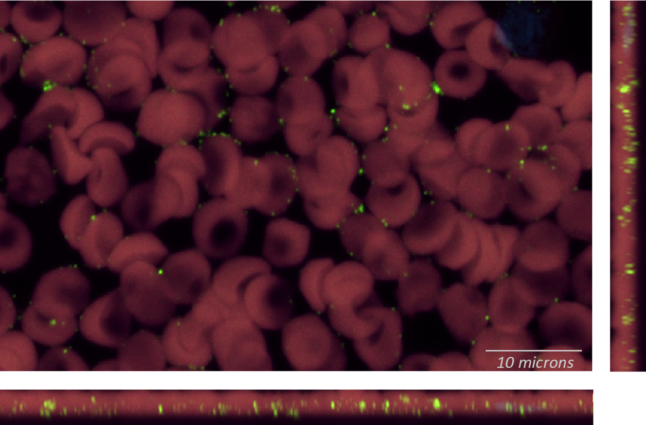
Red Blood Cells
Bartonella henselae RNA
Towards understanding the role of nerve damage in vector-borne diseases.
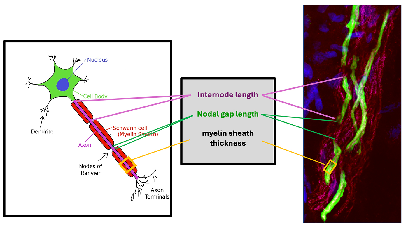
Because neuropathy is one of the most debilitating and challenging to treat symptoms of chronic neurological Lyme, Bartonella and associated diseases, we have developed a new diagnostic protocol to quantify changes in peripheral nerve fibers in biopsies from neuropathic patients with a focus on nerve/pathogen interactions in skin.
We have developed new tools to quantify nerve damage, namely demyelination of nerves in the skin dermis and changes in epidermal nerve density.
We label and image nerves in a skin biopsy and determine if changes have occurred vs. non-affected skin.
Bartonella spp., a probable component of the melanoma pathobiome.
Bartonella bacilliformis (B. bacilliformis), Bartonella henselae (B. henselae), and Bartonella quintana (B. quintana) are bacteria known to cause verruga peruana or bacillary angiomatosis, vascular endothelial growth factor (VEGF)‐dependent cutaneous lesions in humans. Given the bacteria’s association with the dermal niche and clinical suspicion of occult infection by a dermatologist, we determined if patients with melanoma had evidence of Bartonella spp. infection. Within a one‐month period, eight patients previously diagnosed with melanoma volunteered to be tested for evidence of Bartonella spp. exposure/infection. Subsequently, confocal immunohistochemistry and PCR for Bartonella spp. were used to study melanoma tissues from two patients. Blood from seven of the eight patients was either sero-reactive, PCR positive, or positive by both modalities for Bartonella spp. exposure. Subsequently, Bartonella organisms that co‐localized with VEGFC immunoreactivity were visualized using multi‐immunostaining confocal microscopy of thick skin sections from two patients. Using a co‐culture model, B. henselae was observed to enter melanoma cell cytoplasm and resulted in increased vascular endothelial growth factor C (VEGFC) and interleukin 8 (IL‐8) production.
Bartonella henselae Detected in Malignant Melanoma, a Preliminary Study.
Ericson ME, Breitschwerdt EB, Reicherter P, Maxwell C, Maggi RG, Melvin RG, Maluki AH, Bradley JM, Miller JC, Simmons GE Jr. Peterson J, Bae W, Scanlon J, Bemis LTl. Pathogens. 2021; 10(3):326. https://doi.org/10.3390/pathogens10030326
Bartonella spp., a probable component of the melanoma pathobiome.
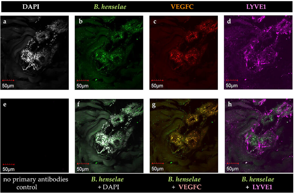
Microscope images show Bartonella henselae in close association with pro‐angiogenic vascular endothelial growth factor C (VEGFC) within dermal lymphatic vessels in melanoma skin biopsy.
Bartonella henselae Detected in Malignant Melanoma, a Preliminary Study.
Ericson ME, Breitschwerdt EB, Reicherter P, Maxwell C, Maggi RG, Melvin RG, Maluki AH, Bradley JM, Miller JC, Simmons GE Jr. Peterson J, Bae W, Scanlon J, Bemis LTl. Pathogens. 2021; 10(3):326. https://doi.org/10.3390/pathogens10030326
Bartonella spp., a probable component of the melanoma pathobiome.

“New Techniques to Diagnose Dangerous Bartonella Infections”
Bartonella henselae Detected in Malignant Melanoma, a Preliminary Study.
Ericson ME, Breitschwerdt EB, Reicherter P, Maxwell C, Maggi RG, Melvin RG, Maluki AH, Bradley JM, Miller JC, Simmons GE Jr. Peterson J, Bae W, Scanlon J, Bemis LTl. Pathogens. 2021; 10(3):326. https://doi.org/10.3390/pathogens10030326
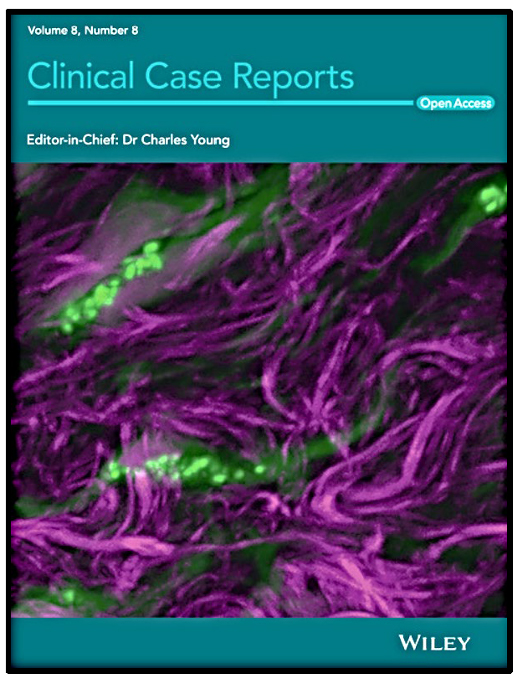
Bartonella biofilm on collagen in skin.
We report on Bartonella bacteria found in apparent biofilm on collagen fibrils in human skin biopsy. The disruption in dermal collagen fiber organization in the non-classical striae-like, skin lesions, Bartonella Tracks, detected using multi-photon (SHG) laser scanning microscopy and immunostaining of Bartonella henselae in patient with Bartonellosis.
Imaging analysis of Bartonella species in the skin using single-photon and multi-photon (second harmonic generation) laser scanning microscopy.
Maluki A, Breitschwerdt E, Bemis L, Greenberg R, Mozayeni BR, Dencklau J, Ericson M. Clin Case Rep. 2020 Aug;8(8):1564-1570. doi: 10.1002/ccr3.2939.
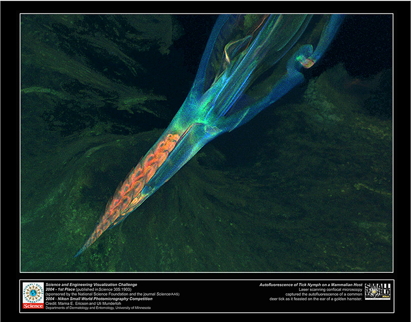
Confocal microscopy image of Ixodes scapularis, the common deer tick on the ear of a golden hamster.
2004 First place in photography in the journal Science/NSF-Science, Engineering, Visualization Challenge.
Marna Ericson, PhD
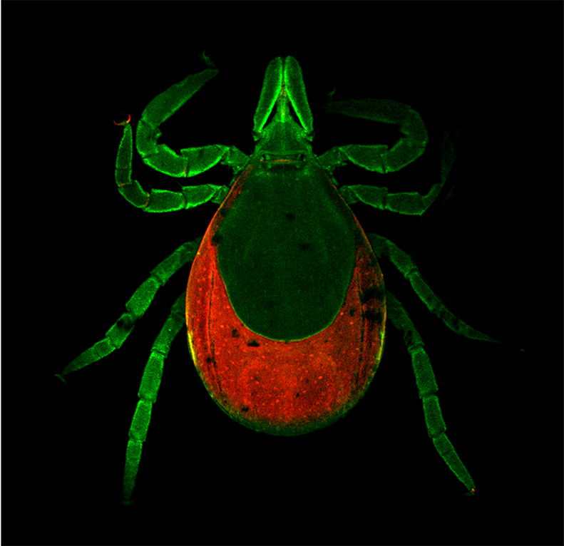
Multi-photon confocal microscopy image of intact Ixodes scapularis, the common deer tick.
2009 semi-finalist in photography in the journal Science/NSF-Science, Engineering, Visualization Challenge.
Marna Ericson, PhD
Collaborate With Us
We are always open to collaborating on potential future research. Please contact us if you’re interested.